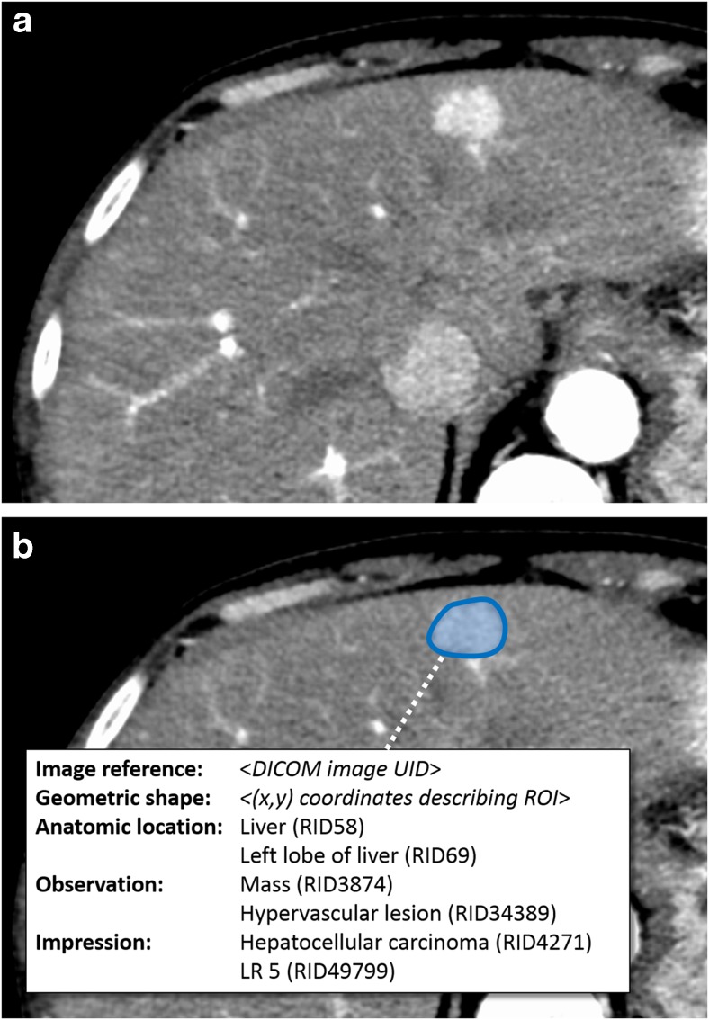
Fig. 1.
Semantic image annotation with AIM. A portion of an axial CT image through the liver after intravenous contrast in arterial phase (a) demonstrates a hypervascular mass in the left hepatic lobe. While a report would describe this lesion, semantic image annotation using the AIM standard (b) enables the findings and diagnosis (white box, inset) to be associated with a specific region of interest (ROI) within the image (blue). AIM uses the extensible markup language (XML) to group a reference to the relevant image (through a DICOM unique identifier, or UID), a geometric description of the ROI, and semantic information such as anatomic location, imaging observations and diagnostic impressions. Such annotations allow the geometric and imaging characteristics of the lesion to be incorporated into other computation. Here for example, the pixel data within the ROI could be quantified to study the enhancement texture characteristics of hepatocellular carcinoma

