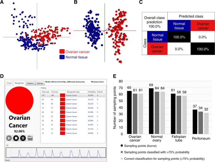
Fig. 4.
Validation of the frozen tissue model using fresh tissue. a 2D PCA model of frozen ovarian cancer samples (>50% tumour content) and all normal samples combined as ‘Normal Tissue’. b 3D LDA component analysis. c Leave-one-patient-out cross-validation of the binary ‘Ovarian Cancer’ vs ‘Normal Tissue’ frozen model, showing 100% correct classification. d OMB Recognition software example showing reported classification for individual sampling points (burns) and the probability value associated with the reported class. e Bar chart showing the numbers of sampling points in fresh tissue classified with >75% probability using the OMB recognition software, plus the diagnostic accuracy at those sampling points [OC 100% (61/61), normal ovary 100% (64/64), fallopian tube 100% (58/58), peritoneum 94.1% (32/34)]

