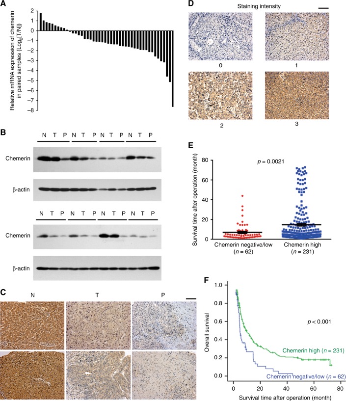
Fig. 1.
Expression pattern and clinical significance of chemerin in HCC. a Expression of chemerin mRNA in 46 pairs of HCC tissues and adjacent non-tumourous liver tissues. b Expression of chemerin proteins in primary HCC tissues (T), portal vein tumour thrombi (P) and paired normal liver tissues (N). c Representative immunohistochemical staining for chemerin in paired normal liver (N), primary HCC (T) and PVTT (P) tissues. Scale bar = 100 µm. d Representative immunohistochemical staining for chemerin with different scores (0, 1, 2, 3) in TMA1. Scale bar = 100 µm. e Scatter diagram reflecting survival of HCC patients and chemerin expression in TMA1. f Kaplan–Meier survival curve of overall survival according to chemerin expression in TMA1, p < 0.001

