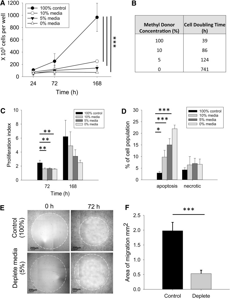
Fig. 2.
Effect of methyl donor depletion on UD-SCC2 cell growth, viability, and migration. a Change in cell number for cells cultured in media containing three different levels of methyl donor depletion (0, 5 and 10%) compared to cells grown in control media (100%). b Cell doubling time for UD-SCC2 cells cultured in media containing three different levels of methyl donor depletion (10, 5 and 0%) compared to cells grown in control media (100%). c Proliferation of cells cultured in media containing three different levels of methyl donor depletion (0, 5, and 10%), compared to cells grown in control media (100%) for 72 and 168 h. Higher proliferation index indicates more proliferating cells and increased proliferation rates. d Proportion of cells undergoing apoptosis (Annexin V positive) or necrosis (PI positive, Annexin V negative) following 168 h depletion (5% depletion; n = 4). e Cell migration into an exclusion zone (white dotted line on representative images) following 72 h depletion was compared to cells grown in complete media (100%). All cells were treated with mitomycin C prior to use in the migration assay to inhibit cell proliferation. f Area of migration quantified from three independent experiments (independent Student’s t test). One-way independent ANOVA with Bonferroni post-hoc comparison; *p < 0.05, **p < 0.01, **p < 0.005

