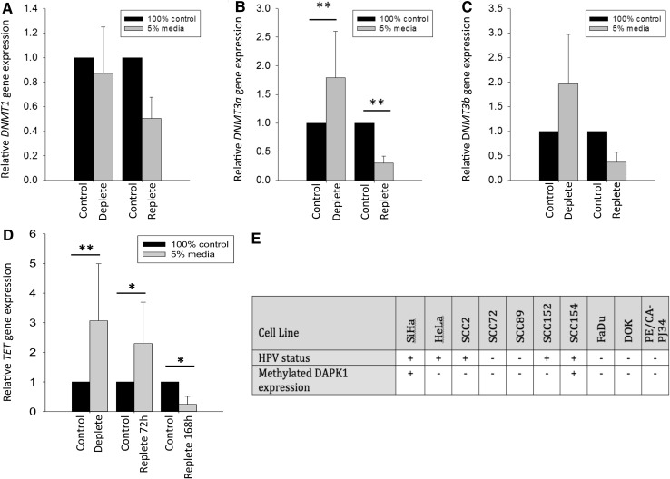
Fig. 4.
Effect of methyl donor depletion and repletion on DNMT and TET1 expression. Gene expression of DNMT1 (a), DNMT3a (b), and DNMT3b (c) and d TET1 relative to β2 M expression was measured using qPCR in UD-SCC2 cells. Expression was measured following methyl donor depletion for 168 h with medium containing 5% methyl donors (deplete) followed by a further 72 h culture in medium containing 100% methyl donors (and 168 h for TET1) (replete). Control cells were continuously cultured in 100% methyl donor containing medium alongside deplete and replete cells. n = 6, independent Student’s t test, **p < 0.01. e Presence of methylated DAPK1 promoter in a panel of HNSCC cell lines as measured by qMSP; the cervical cell lines SiHa were used as controls. Table shows cells positive for DAPK1 methylation (+, Ct values <35) and cells negative for methylated DAPK1 (−, Ct values >35). All cell lines tested were positive for β-actin (mean Ct value 25 ± 0.47)

