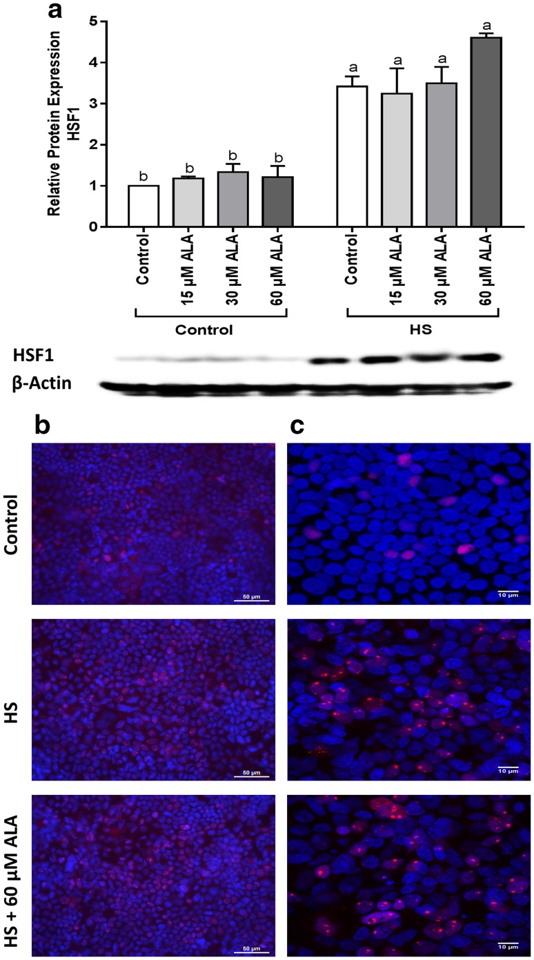
Fig. 1.
HS upregulates the HSF1 protein expression and induces HSF1 nuclear granules. Caco-2 cells grown on inserts and pretreated with ALA (24 h) were exposed to HS (42 °C, 6 h). HSF1 protein expression (normalized with β-actin) relative to unstimulated cells evaluated by WB analysis (a), is expressed as mean ± SEM of three independent experiments. Different lower case letters denote significant differences among groups. Localization of HSF1 was visualized by immunofluorescence staining. Objective ×40 (b) and ×63 (c)

