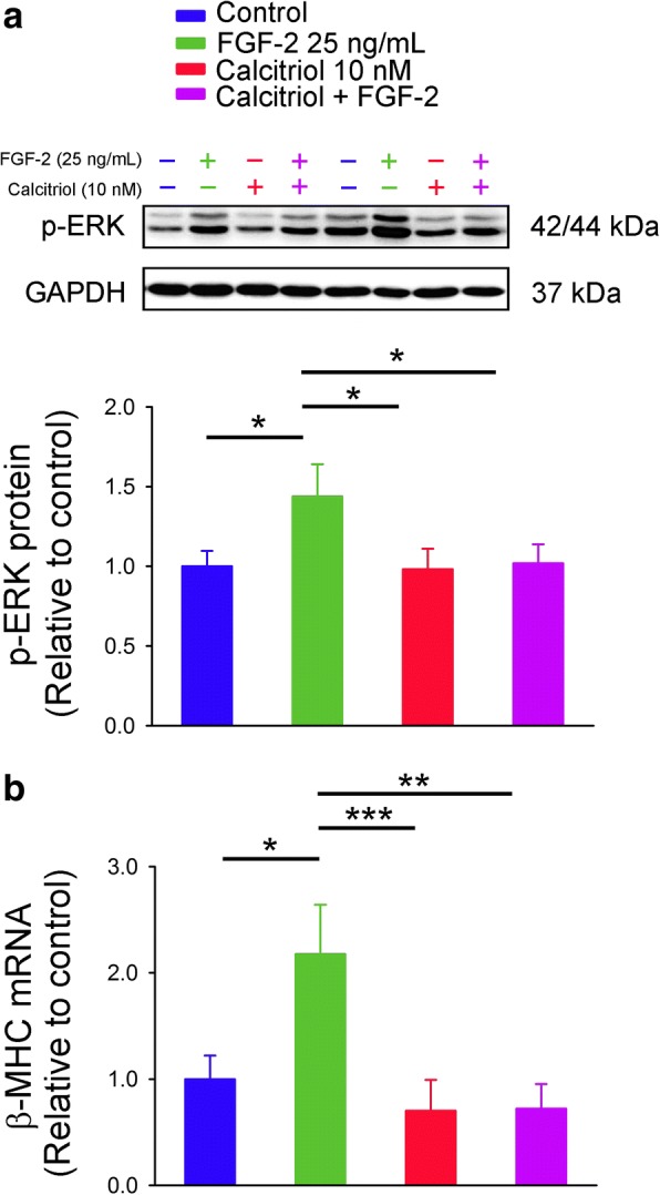
Fig. 2.

Phosphorylated extracellular signal-regulated kinase (p-ERK) protein and β-myosin heavy chain (β-MHC) mRNA expressions in control and calcitriol-treated HL-1 cells without and with fibroblast growth factor (FGF)-2 incubation. (a) FGF-2 (25 ng/mL) was administered for 30 min to study ERK phosphorylation (n = 6), or (b) for 48 h to evaluate β-MHC mRNA from control and calcitriol (10 nM)-treated HL-1 cells (n = 6). * p < 0.05, ** p < 0.01, *** p < 0.005
