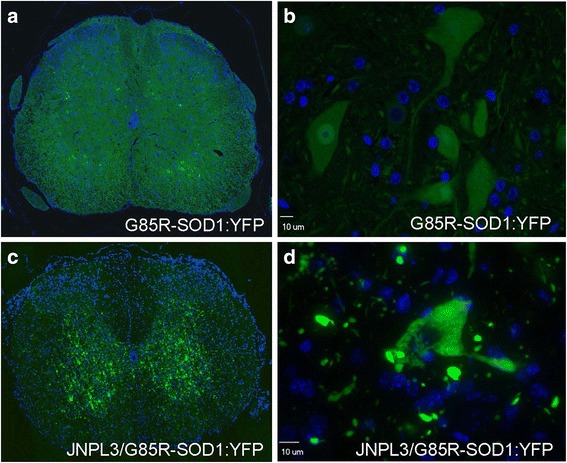
Fig. 1.

G85R-SOD1:YFP aggregation into punctate inclusions within the spinal cord of JNPL3/G85R-SOD1:YFP mice. Compared to the diffuse distribution of G85R-SOD1:YFP in spinal motor neurons of single transgenic animals (a and b), the fluorescence is organized into large neuropil inclusions with granular/punctate accumulation in the cell bodies of spinal motor neurons of bigenic JNPL3/G85R-SOD1:YFP mice (c and d). Exposure times were kept consistent across images and set to capture images of the inclusions in the bigenic mice at optimal exposure. Nuclei were stained with DAPI (blue). All animals analyzed were female. Representative images (40× magnification) of the ventral horn within the spinal cord are shown for 8 JNPL3-G85R-SOD1:YFP double transgenic mice and 3 G85R-SOD1:YFP single transgenic mice (aged 7 to 15.5 months). An additional low power image of the spinal ventral horn bigenic JNPL3/G85-SOD1:YFP mice is provided in Additional file 2: Figure S2a
