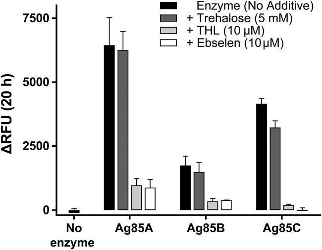
Fig. 2.
Fluorescence resulting from QTF exposure to the native M. tuberculosis mycolyltransferases Ag85A, Ag85B, and Ag85C. Fluorescence emission was measured after incubation of QTF (1 μM) with purified M. tuberculosis Ag85A, Ag85B, or Ag85C (20 nM) for 20 h at 37 °C in 20 mM phosphate buffer (pH = 7.2) (black bars). Turnover was also assessed in the presence of a mycolyl acceptor (5 mM trehalose, dark gray bars) and generic mycolyltransferase inhibitors tetrahydrolipstatin (THL) (10 μM light gray) or ebselen (10 μM white). RFU, relative fluorescence units. Error bars depict SD.

