Fig. 1.
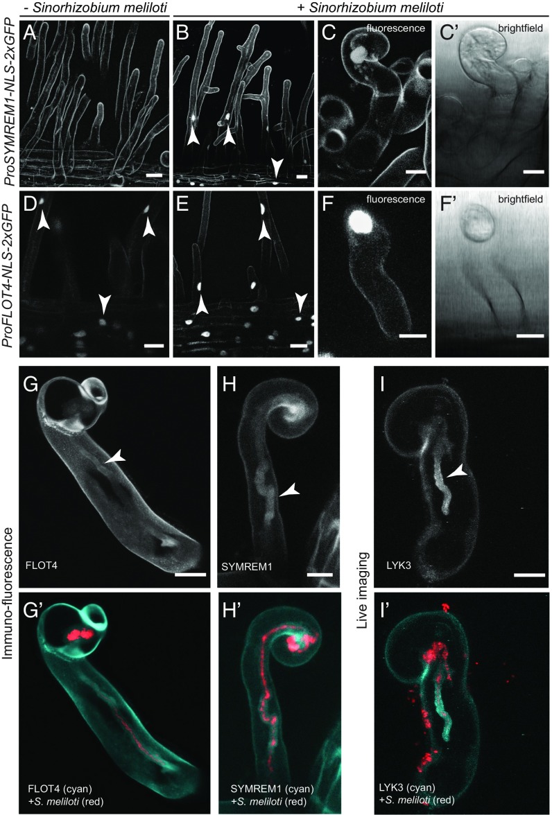
Expression and localization analyses of SYMREM1 and FLOT4. Monitoring promoter activation of SYMREM1 (A–C′) and FLOT4 (D–F′) with cellular resolution in transgenic M. truncatula root hairs using the genetically encoded reporters ProSYMREM1:NLS:2xGFP and ProFLOT4:NLS:2xGFP, respectively. (A and B) Activation of SYMREM1 as indicated by nuclear fluorescence (arrowheads) in uninoculated (A) and S. meliloti 2011 (mCherry) inoculated (B) roots. (C, C′, F, and F′) Close-ups of individual curled root hairs with fluorescent nuclei. (A–C′) Autofluorescent contours of roots hairs and epidermal cells are visible due to ultra-sensitive imaging settings that were chosen due to low signal intensities. (D–F′) Activation of the FLOT4 promoter in uninoculated (D) and inoculated (E) conditions (arrowheads). (G–H′) Immunofluorescence targeting FLOT4-GFP (G and G′) and GFP-SYMREM1 (H and H′), both expressed from their endogenous promoters. (I and I′) Transgenic line expressing a ProLYK3::LYK3-GFP construct. Arrowheads indicate the localization of the respective protein around the ITs. (Scale bars: 20 µm in A, B, D, and E; 10 µm in C, C′, F, F′, G, H, and I.)

