Fig. 3.
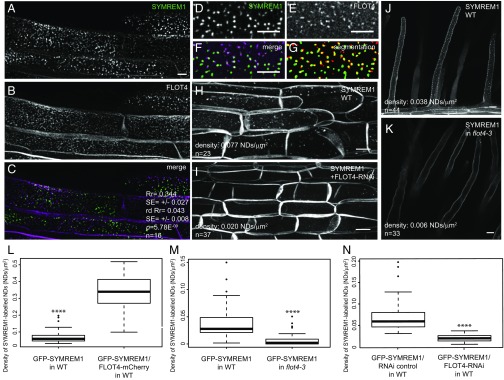
SYMREM1 is recruited to nanodomains (NDs) in a FLOT4-dependent manner. (A–C) GFP-SYMREM1 (A) and FLOT4-mCherry (B) colocalized (C) in transgenic M. truncatula root epidermal cells. Quantification data are depicted within images. Rr, Pearson correlation coefficient; rdRr, Pearson correlation coefficient obtained after image randomization of the FLOT4-mCherry channel; P, confidence interval obtained from a Student t test comparing Rr and rdRr with the number of replicates (n) indicated in the panels. (D and E) Close-up views of nanodomain-localized GFP-SYMREM1 (D) and FLOT4-Cherry (E) at the cell surface. (F and G) Colocalization was quantified in merged images (F) and after image segmentation (G). (H–J) Density of GFP-SYMREM1-labeled nanodomains was greatly reduced on coexpression with a FLOT4-RNAi construct in root epidermal cells (I) and in flot4-3 mutant root hairs (K) compared with the respective controls (H and J). (L) Density of GFP-SYMREM1–labeled nanodomains was significantly increased when coexpressed with FLOT4-mCherry. (M and N) Quantification on GFP-SYMREM1–labeled nanodomains in the flot4-3 mutant (M) and FLOT4-RNAi (N) roots. ****P < 0.0001, Student t test. (Scale bars: 5 μm in A–F; 10 μm in H–K.)

