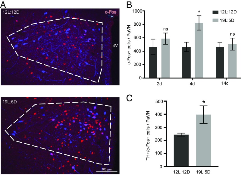
Fig. 1.
Activity of PaVN neurons is elevated after long-day photoperiod exposure. WT rats were exposed to either a long-day photoperiod (19L:5D) or balanced-day photoperiod (12L:12D) for 2, 4, or 14 d. Immunofluorescent staining of TH and c-Fos was performed with fixed brain sections. (A) Confocal images of the PaVN after 4 d of exposure; white dashed lines indicate the PaVN boundary. 3V, third ventricle. (B) Quantification of the number of c-Fos+ cells in the PaVN per animal after different durations of exposure: 12L:12D for 2 d, n = 4 animals; 19L:5D for 2 d, n = 4 animals; 12L:12D for 4 d, n = 6 animals; 19L:5D for 4 d, n = 6 animals; 12L:12D for 14 d, n = 4 animals; and 19L:5D, n = 5 animals. Welch’s t test (2 d, P = 0.4267; 4 d, P = 0.0216; 14 d, P = 0.7209). Data are mean ± SEM. *P < 0.05. ns, not significant. (C) Quantification of the number of TH+/c-Fos+ cells in the PaVN per animal after 4 d of exposure to 12L:12D or 19L:5D (n = 6 animals per condition). Welch’s t test (P = 0.0478). Data are mean ± SEM. *P < 0.05.

