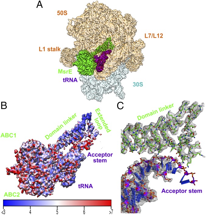
Fig. 1.
Cryo-EM structure of the MsrE-ribosome complex. (A) Electron density of the overall complex. Ribosome 50S and 30S subunits are shown in pale orange and cyan, respectively. The electron density of 50S is made partially transparent to reveal the densities corresponding to MsrE protein (green) and tRNA (purple). (B) Local resolution of MsrE and tRNA shown in same orientation as in A. (C) Cryo-EM electron density of the MsrE domain linker extended loop and tRNA acceptor stem region.

