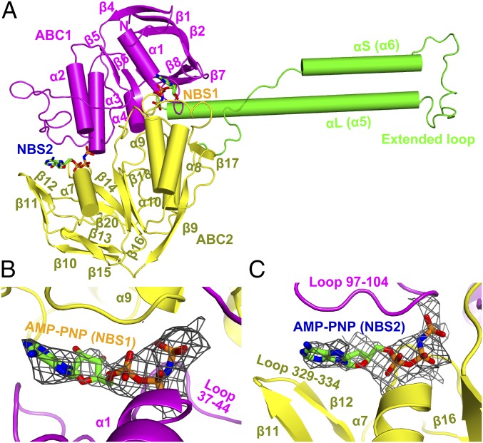
Fig. 2.
Structure of the ribosome-bound MsrE protein. (A) MsrE protein shown in cartoon with secondary structure elements of ABC1 (magenta), ABC2 (yellow), and domain linker (green) labeled. The nonhydrolyzable ATP analog AMP-PNP is shown bound to NBSs NBS1 and NBS2. (B and C) Close-up views of the MsrE NBS1 (B) and NBS2 (C) with the cryo-EM electron density map shown for AMP-PNP.

