Fig. 5.
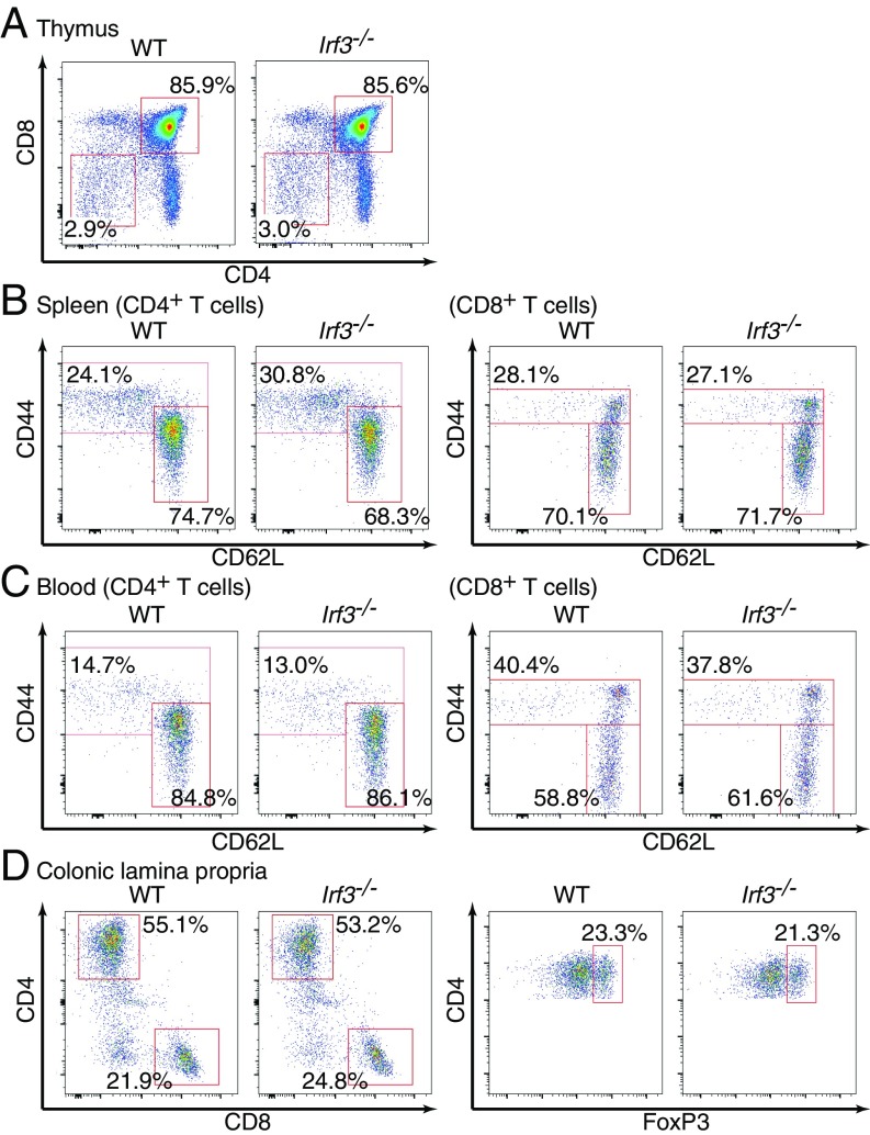
Normal T cell subsets in Irf3-deficient mice. (A) Thymocytes from WT or Irf3–/– mice were analyzed by flow cytometry. Percentages of CD4–CD8– and CD4+CD8+ cells in the total thymocytes are shown. (B) Splenocytes were prepared from WT or Irf3–/– mice and analyzed by flow cytometry. CD4+TCRβ+ T cells (Left) and CD8+TCRβ+ T cells (Right) were gated. Percentages of effector/memory T cells and naïve T cells in the gated population are shown. (C) Whole-blood cells were prepared from WT or Irf3–/– mice. Red blood cells were lysed, and the remaining cells were analyzed by flow cytometry as in B. (D) Colonic lamina propria cells were obtained from WT or Irf3–/– mice and subjected to flow cytometry analysis. TCRβ+ T cells (Left) and CD4+TCRβ+ T cells (Right) were gated. The results shown are representative of three independent experiments.

