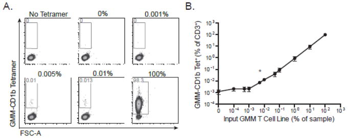
Figure 3. Quantitative validation of the limit of detection of the CD1 tetramer assay.
The GMM-specific T-cell line was serially diluted with CD1d-restricted T-cells specific for α-GalCer and then stained with GMM-CD1b tetramer. (A) Representative staining from one experiment in which GMM T cells were diluted with CD1d-restricted T ranging from no GMM-specific T-cells (0%) up to only GMM-specific T-cells (100%). ‘No Tetramer’ refects the condition in which no tetramer was added to a 1:1 mixture of GMM T cells and α-GalCer T cells). (B) Summary analysis of three independent experiments. Data are depicted as a percent of total CD3+ cells. Shown are the mean and standard error of the mean at each dilution. Chi-square analysis demonstrates that 0.007% is the limit of detection of the assay (marked with an asterisk).

