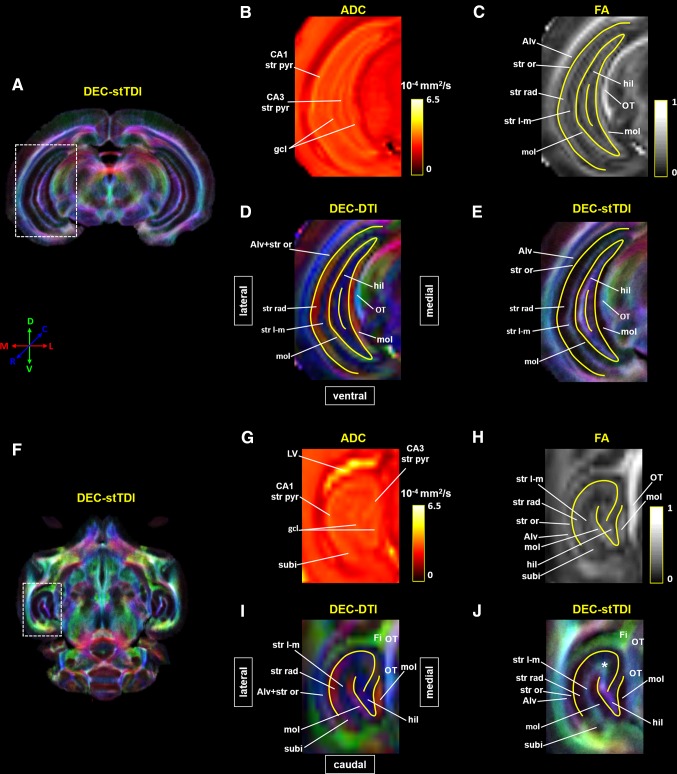
Fig. 3.
Laminar organization of tree shrew hippocampal formation (Hipp) revealed by DTI and stTDI. A, F Super-resolution DEC-stTDI maps of a coronal slice and a transverse slice at the level of the Hipp. The hippocampal areas (dashed white rectangles) are enlarged in E and J. Native-resolution apparent diffusion coefficient (ADC) (B, G), fractional anisotropic (FA) (C, H), and DEC-DTI (D, I) maps of the corresponding areas are also shown. Yellow solid lines in C–E and H–J were manually drawn according to the ADC contrast to show the location of the granule cell layer (gcl) of the dentate gyrus and the stratum pyramidale (str pyr) of CA1 and CA3. The white asterisk in J indicates a structure revealed by the stTDI reconstruction only. Alv alveus, Fi fimbria of hippocampal formation, gcl granule cell layer of the dentate gyrus, hil hilar region of the dentate gyrus, LV lateral ventricle, mol molecular layer of the dentate gyrus, OT optic tract, str l-m stratum lacunosum-moleculare, str or stratum oriens, str pyr stratum pyramidale, str rad stratum radiatum, subi subiculum.

