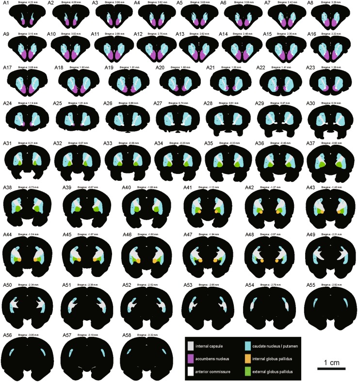
Fig. 2.
Schematic drawings showing the general morphology of the striatum and globus pallidus in the coronal plane from rostral to caudal in the tree shrew (A1-A58, referring to The Tree Shrew (Tupaia belangeri chinensis) Brain in Stereotaxic Coordinates [52]). Colors represent distinct subregions of the basal ganglia. ICj, islands of Calleja. Scale bar, 1 cm.

