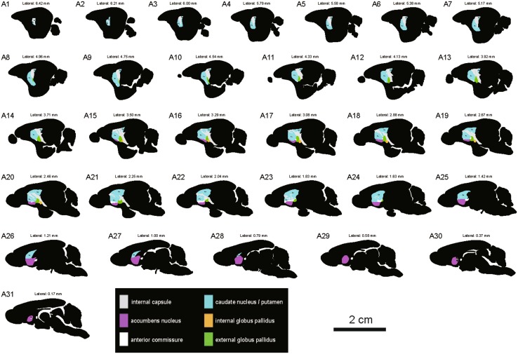
Fig. 5.
Schematic drawings showing the general morphology of the striatum and globus pallidus in the sagittal plane from lateral to medial in the tree shrew (A1–A31, referring to The Tree Shrew (Tupaia belangeri chinensis) Brain in Stereotaxic Coordinates [52]). Colors represent distinct subregions of the basal ganglia. ICj, islands of Calleja. Scale bar, 2 cm.

