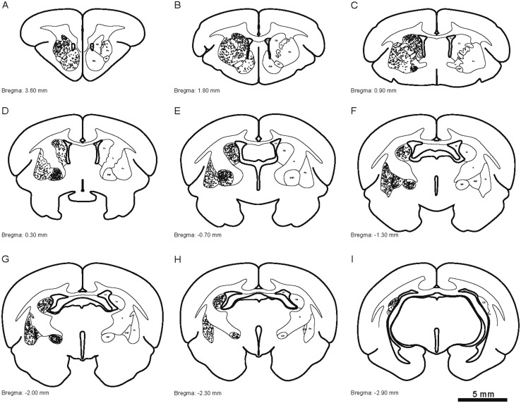
Fig. 8.
Camera lucida drawings showing the distribution of parvalbumin-ir cells (dots) in the striatum and globus pallidus of the tree shrew from rostral to caudal (A–I). Representative coronal sections were selected, with reference to bregma. The density of dots represents the relative density of cells in the areas. Each dot represents approximately one parvalbumin-labeled neuron. ac, anterior commissure; Acb, accumbens nucleus; Cd, caudate nucleus; EGP, external globus pallidus; ic, internal capsule; IGP, internal globus pallidus; LV, lateral ventricle; Pu, putamen.

