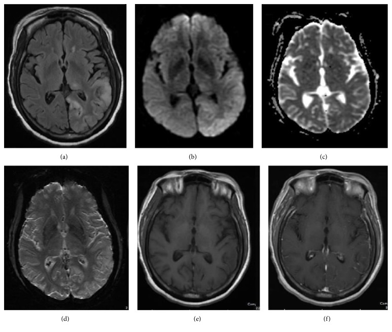
Figure 1.
64-year-old female with confusion, right lower extremity weakness, and questionable encephalitis. Axial FLAIR MR image (a) shows increased signal intensity and gyriform swelling in the cortex and subcortical white matter of the left parietooccipital lobe and a focus in the left thalamus. Diffusion weighed (b) and attenuation diffusion coefficient, ADC, MR images (c) reveal mild decreased diffusivity. No susceptibility artifact or contrast enhancement is observed on axial susceptibility weighted image, SWI (d), and axial T1 weighted (e) and axial T1 weighted postgadolinium images (f), respectively.

