
Fig. 3.

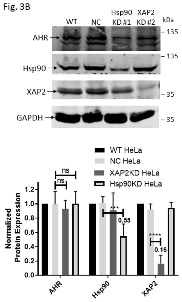
Down-regulation of the Hsp90 and XAP2 proteins did not decrease the AHR protein levels in HeLa cells. (A) AHR protein levels were determined by Western analysis in Hsp90- (left panel) and XAP2- (right panel) knockdown stable HeLa cells. Eight cell lines were examined: wild type (WT), negative control knockdown stable (NC), Hsp90 knockdown (KD) #1–3, 5, 6, and XAP2 knockdown (KD) #3. The images in left panel were performed once whereas images in right panel were repeated with different samples twice with similar results (n=3). (B) Western analysis of AHR, Hsp90, and XAP2 expression in wild-type HeLa cells (WT), scramble shRNA-treated stable HeLa cells (NC), Hsp90-knockdown stable HeLa cells using #1 shRNA (Hsp90KD), and XAP2-knockdown stable HeLa cells using #2 shRNA (XAP2KD). Images on top are representative of the replicates and the graph was plotted below (n = 6, means ± SD). Each lane of A and B contained 40 μg of whole cell extract and was normalized by GAPDH.