
Fig. 6.

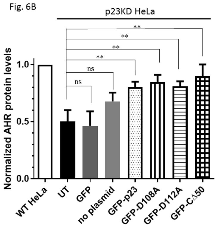
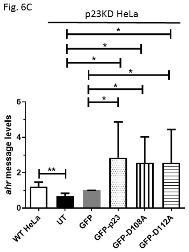

GFP fusion of wild type p23 and p23 mutants restored the AHR protein and message levels in p23 knockdown (p23KD) stable HeLa cells. (A) Western results showing GFP-p23 wild type (WT), GFP-p23 D108A, GFP-p23 D112A, and GFP-p23 CΔ50 restored the AHR protein levels in p23KD HeLa cells. UT, untransfected p23KD cells; no plasmid, p23KD cells had undergone transfection with no plasmid; GFP, p23KD transfected with the GFP control plasmid. The image is representative of triplicate data. Each lane contained 40μg of protein. (B) Quantitative analysis of western blot in A. (C) Exogenous p23 treatment also restored the ahr message levels in p23KD HeLa cells. The plots are quantified qPCR data showing the means with error bars (mean ± SD, n=5 for all except n=7 for GFP and GFP-p23 D112A groups). Statistical significance were calculated when compared to untransfected (UT) p23KD cells or p23KD cells transfected with the GFP control plasmid. (D) Florescence microscopy using a Keyence microscope showing that GFP-p23 WT was nuclear whereas p23 mutants (D108A and D112A) were cytoplasmic and nuclear.