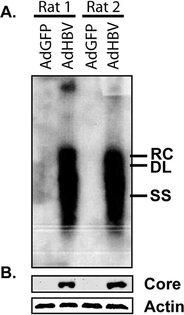
Fig 2. Confirmation of HBV replication.
HBV replication was monitored by Southern blot analysis of HBV core particle-associated DNA (A) and western blot for HBV core protein (B). For western blot analysis, β–actin was used as a protein loading control. Samples were collected at 72 hr (48 hr after infection). RC – relaxed circular DNA, DL – double-stranded linear DNA, SS – single stranded DNA.

