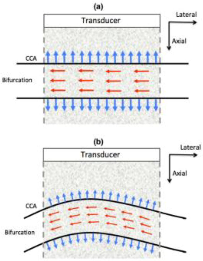
Fig. 1.
Illustrations showing examples of the orientation of the carotid artery relative to the ultrasound transducer. The red and blue arrows indicate the direction of blood flow and the radial deformation in the vessel wall, respectively. The figures correspond to the instances when the radial deformation in the vessel wall is (a) aligned and (b) not aligned with the direction of beam propagation (axial).

