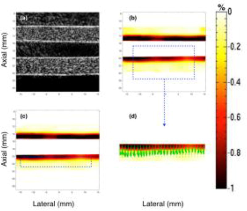
Fig. 4.
Montage of ultrasound sonogram (a), principal (b) and axial (c) strain elastograms, and the direction vectors map (d) associated with the major component of the principal strain displayed in (b). These images are obtained from a homogenous vessel phantom at a transducer angle of 0°, using CPW imaging.

