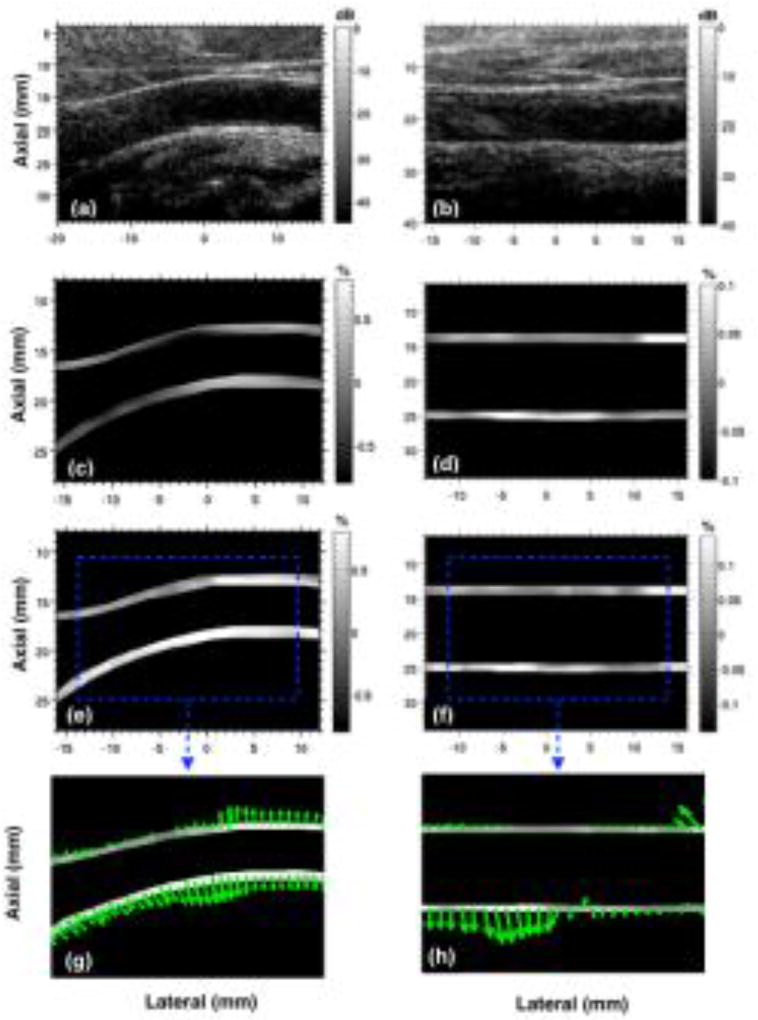
Fig. 7.
Montage displays representative images of carotid artery sonogram (a,b) and their corresponding axial (c,d) and principal (e,f) strain elastograms associated with two healthy male subjects. Subplots (g,h) corresponds to the principal strain direction vectors associated with (e,f). The two columns correspond to images from two healthy volunteers with their common carotid artery non-parallel (a,c,e) and parallel (b,d,f) to the transducer.

