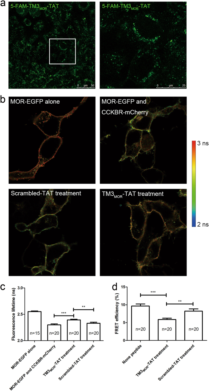
Fig. 4. TM3MOR-TAT peptide disrupted MOR–CCKBR interaction.
a An example of confocal microscopy image of 5-FAM-labeled TM3MOR-TAT-treated HEK293 cells showing successful penetration of the TM3MOR-TAT peptides into the cells (left). The magnified area indicates the membrane location of the peptide (right). b An example of the FLIM image of TAT-fused peptide-treated cells co-expressing MOR-EGFP and CCKBR-mCherry. Compared with the untreated or scrambled-TAT-treated group, EGFP in the TM3MOR-TAT-treated cells showed longer lifetime of fluorescence. c Statistics of EGFP fluorescence lifetime in (b). d FRET efficiency of MOR-EGFP and CCKBR-mCherry in interfering peptide-treated cells. Compared with the non-peptide-treated or scrambled-TAT-treated group, the TM3MOR-TAT-treated group exhibited a significant decrease in FRET efficiency. *p < 0.05, **p < 0.01, t-test. Data are represented as the mean ± SEM

