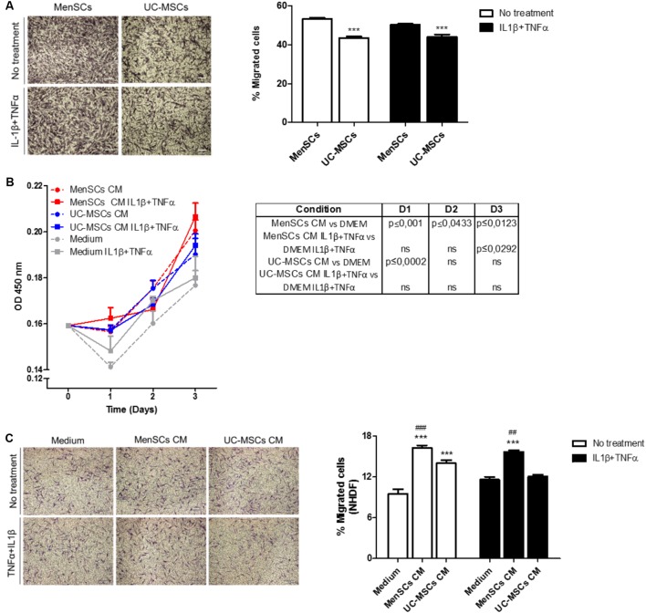
FIGURE 3.
MenSCs and MenSCs-secretome exhibits increased migratory effects. (A) Migration of MenSCs and UC-MSCs with and without proinflammatory stimuli. Migration was evaluated by the Transwell assay system. Left panel, representative images of migrated cells. Right panel, quantification of the area of migrated cells per photographic field using Image J software. (B) Effect of MSCs-CM on NHDF proliferation. MSCs-CM was prepared by incubating MSCs (2 × 10⁵/well) in serum-free medium or with IL1β and TNFα (10 ng/ml each). Right, table with statistical analysis comparing MSCs-CM vs. Medium. (C) Effect of MSCs-CM on NHDF migration. Migration was evaluated by Transwell assay system. MSCs-CM was prepared by incubating MSCs (2 × 10⁵/well) in serum-free medium or with IL1β and TNFα (10 ng/ml each). Left panel, representative images of migrated cells. Right panel, quantification of the area of migrated cells per photographic field using Image J software. Values are expressed as the mean ± SE of at least 3 independent experiments in triplicates. For (A) UC-MSCs vs. MenSCs ∗∗∗p ≤ 0.001 and for (C) MSCs-CM vs. Medium, ∗∗∗p ≤ 0.001; MenSCs-CM vs. UC-MSCs CM, ###p ≤ 0.001, ##p ≤ 0.01. CM, conditioned medium; MenSCs, menstrual derived mesenchymal stem cells; UC-MSCs, umbilical cord mesenchymal stem cells; NHDF, normal human dermal fibroblast; TNFα, tumor necrosis factor alpha; IL1β, interleukin 1 beta.

