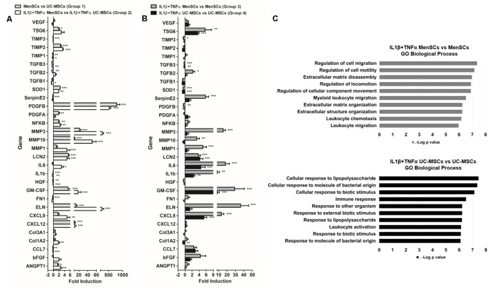
FIGURE 4.
MenSCs exhibited a robust wound healing transcriptomic pattern. MSCs were cultured in basal conditions (medium serum free) and with pro-inflammatory cytokines, IL1β and TNFα (10 ng/ml each) during 24 h. The cells were recovered and total mRNA was extracted, reverse-transcribed and RT-qPCR was performed using Stratagene Mx3000P. The quantitative data from the RT-PCR was normalized by the expression of GAPDH. Expression of different genes involved in wound healing process, (A) fold induction of MenSCs vs. UC-MSCs and IL1β+TNFα MenSCs vs. IL1β+TNFα UC-MSCs and (B) fold induction of IL1β+TNFα MenSCs vs. MenSCs and IL1β+TNFα UC-MSCs vs. UC-MSCs. Values are expressed as the mean ± SE of at least 3 independent experiments in triplicates. ∗p ≤ 0.05, ∗∗p ≤ 0.01, ∗∗∗p ≤ 0.001 compared to their corresponding MSCs condition. (C) GO analysis showing the 10 most important biological process of IL1β+TNFα MenSCs vs. MenSCs and IL1β+TNFα UC-MSCs vs. UC-MSCs. MSCs, mesenchymal stem cells; MenSCs, menstrual derived mesenchymal stem cells; UC-MSCs, umbilical cord-derived mesenchymal stem cells; TNFα, tumor necrosis factor alpha; IL1β, interleukin 1 beta.

