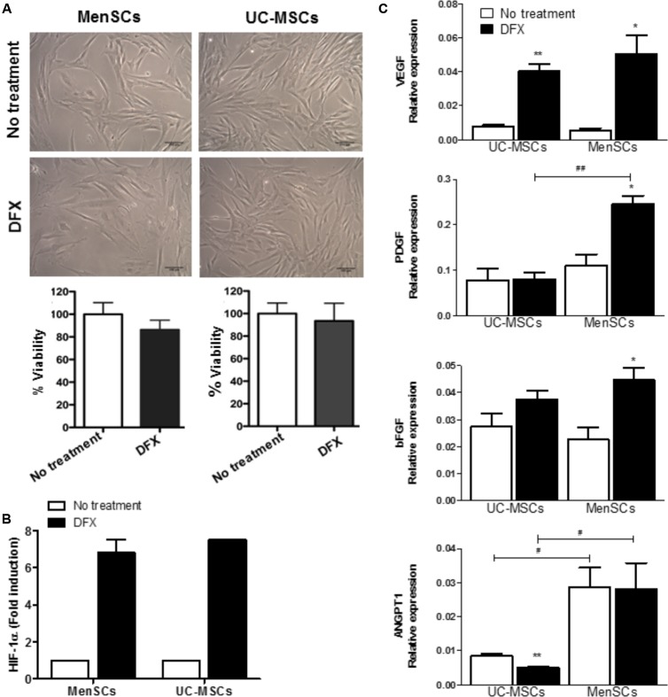
FIGURE 5.
Increased expression levels of pro-angiogenic factors in hypoxia conditions. MSCs were incubated during 24 h with 150 μM of DFX. (A) Upper: Representative images showing no changes of morphological characteristics of MSCs comparing to no treated control (without DFX). Lower: Percentage of MSCs viability quantified by trypan blue exclusion assay. (B) Levels of HIF-1α determined by ELISA in MSCs lysates. Data were expressed as fold change comparing to non-treated control (without DFX). Values are expressed as the mean ± SE. (C) Expression of mRNA levels of the pro-angiogenic factors VEGFα, ANGPT1, PDGF, and bFGF. Values are expressed as the mean ± SE. Treated DFX MSCs vs. non-treated MSCs, ∗p ≤ 0.05, ∗∗p ≤ 0.01. MenSCs vs. their respective condition in UC-MSCs, #p ≤ 0.05, ##p ≤ 0.01. MSCs, mesenchymal stem cells; MenSCs, menstrual derived mesenchymal stem cells; UC-MSCs, umbilical cord-derived mesenchymal stem cells; DFX, deferoxamine; HIF-1α, hypoxia inducible factor 1 alpha; VEGF, vascular endothelial growth factor; ANGPT1, angiopoietin 1; PDGF, platelet derived growth factor; bFGF, basic fibroblast growth factor.

