
Figure 1.
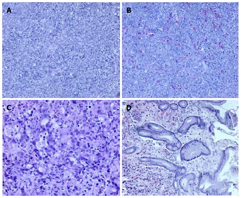
Histological ovary assay. A: Signet ring of tumor cell infiltration within the ovarian stroma (10 ×, HE); B: Mucicarmine staining highlights the presence of mucin in the cytoplasm (10 ×). Histopathologic evaluation of antral mucosa showing (C) poorly differentiated carcinoma infiltration with signet ring cells (20 ×, HE) and (D) mucicarmine staining of mucin-positive cells in the gastric mucosa (10 ×).