
Fig. 6. Relative phosphorylation of AMPK in hepatic cell (HepG2) upon incubation with different treatments for 3 h at 37°C under 5% CO2.
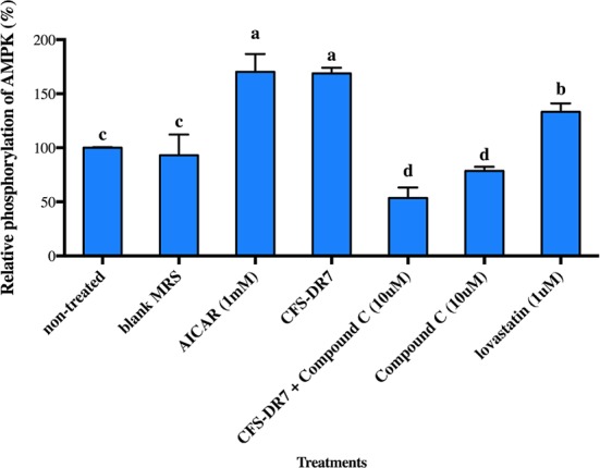
Results are expressed as mean; error bars (SEM); n=3. Statistical analysis was performed using one-way ANOVA with Duncan’s post hoc comparisons. Means labelled with different letters are significantly different (p<0.05). AICAR (AMPK activator) and Compound C (AMPK inhibitor) were used as positive and negative control, respectively. Non-treated: HepG2 cultured in serum-free Dulbecco's modified Eagle's medium (DMEM) without any treatment; blank MRS, HepG2 cultured in sterile de Mann, Rogosa, Sharpe (MRS) broth at a final concentration of 30% (v/v) in serum-free DMEM; CFS-DR7, HepG2 cultured in CFS from Lactobacillus plantarum DR7 at a final concentration of 30% (v/v) in serum-free DMEM.