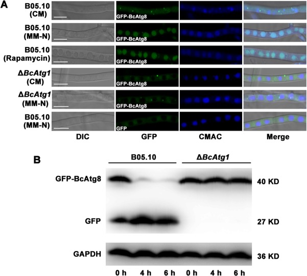
FIG 3.
Analysis of autophagy process using GFP-BcAtg8 marker in B. cinerea. (A) GFP-BcAtg8 localized in the cytoplasm as preautophagosomal structures (PAS) under nutrient-rich conditions; with induction by starvation or rapamycin, the autophagy process was activated and GFP-BcAtg8 transferred to vacuoles. The vacuoles were stained with CMAC (7-amino-4-chloromethylcoumarin) and examined by fluorescence microscopy. DIC, differential interference contrast; CM, complete medium; MM-N, minimal medium without (NH4)2SO4. Scale bars, 10 μm. (B) GFP-BcAtg8 proteolysis assays of B05.10 and the ΔBcAtg1 mutant. Mycelia were cultured at 25°C for 48 h in CM liquid medium, and autophagy was induced after 4 or 6 h of nitrogen starvation. Mycelia were collected at the indicated times, and mycelial extracts were analyzed by anti-GFP antibody Western blotting. GAPDH was used as an internal reference.

