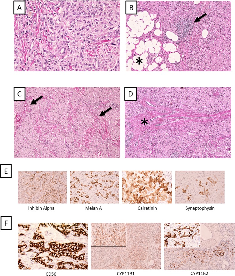
Figure 2.
Histology of extensive TARTs in a man with 3βHSD2D. (A) Routine hematoxylin and eosin staining of the left TART. The tumor cells have a large nuclear-to-cytoplasmic ratio, and tumor nuclei are round to elliptical with a loose chromatin. The cytoplasm is eosinophilic and granulated. (B) Associated TART features such as adipose tissue metaplasia (asterisk) and focal lymphocytic infiltrates (arrow) are noted. (C) The tumor is seen with a massive peritubular fibrosis and hyalinization (arrow). (D) Tumor within the rete testis (asterisk), the hypothesized origin of TARTs. (E) Immunohistochemical markers for adrenal tissue; from left to right: inhibin A, Melan A, calretinin, and synaptophysin. (F) Additional immunohistochemical markers (CD56, CYP11B1, and CYPP11B2) supporting the TART diagnosis. Widespread CD56 immunoreactivity and focal CYP11B1 and CYP11B2 positivity. All photomicrographs are magnified ×100, except A and inserts of CYP11B1/2 immunostainings (×400).

