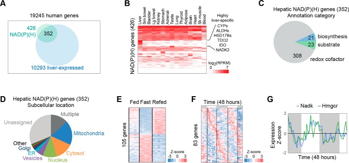
Figure 3.
Compartmentalization of the hepatic NAD(P)ome. A, hepatic NAD(P)ome is defined as the 352 members of the human NAD(P)ome that show expression in liver. B, tissue expression is shown across 15 human tissues selected from the GTEx atlas. Expression is shown as RPKM mapped reads. C, relative distribution of hepatic enzymes using NAD(P)+ as cofactors in redox reactions, as substrates, or in NAD(P)+ biosynthesis. D, subcellular distribution of the hepatic NAD(P)ome with data available from the Human Protein Atlas (HPA). E, temporal compartmentalization during fasting/feeding is shown for 105 hepatic NADPome genes showing significant differential expression changes based on the Montagner et al. (54) study in the mouse (Student's t test p value < 0.05 after Bonferroni correction). F, gene expression is shown for the 83 genes showing significant circadian periodicity from Hughes et al. (57). Gray bars indicate periods of darkness. G, circadian expression of NADK and HMGCR based on data from Hughes et al. (57).

