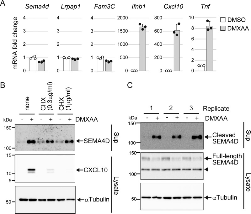
Figure 3.
Activation of STING induces shedding of SEMA4D in a transcription-independent manner. A and C, Raw264.7 cells were cultured in 0.1% FCS/DMEM containing DMSO (−) or 100 μg/ml DMXAA for 4 h. In A, the mRNA levels of the indicated genes were determined by real-time PCR. The data are normalized by β-actin mRNA and shown as -fold change relative to DMSO control. Scatter plots show the individual data, and bar graphs indicate mean ± S.D. (n = 3). B, Raw264.7 cells were cultured in 0.1% FCS/DMEM, with or without the indicated concentrations of CHX for 30 min, and then further incubated with DMSO (−) or 100 μg/ml DMXAA for 4 h. SEMA4D and CXCL10 levels in the culture supernatants and the α-tubulin level in cell lysates were determined by Western blotting with anti-SEMA4D, anti-CXCL10, and anti-α-tubulin. In C, the protein levels of soluble SEMA4D in culture supernatants and membrane SEMA4D in cell lysates from three biological replicates were analyzed by Western blotting with anti-SEMA4D. The arrowhead indicates an intracellular cleaved form of SEMA4D.

