Figure 3.
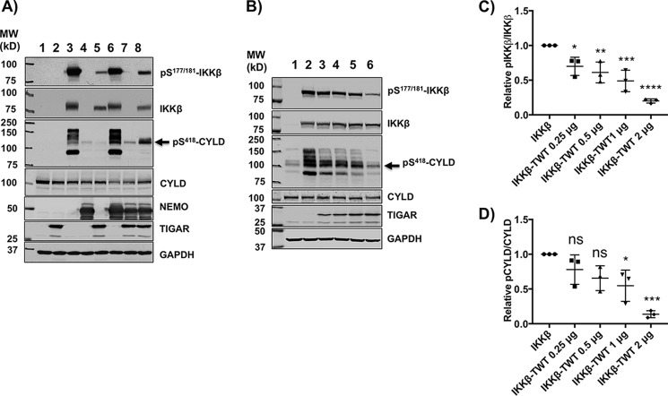
TIGAR inhibits IKKβ-dependent phosphorylation of several direct IKKβ cellular substrate targets. A, HEK293T cells were transfected with the indicated combinations of cDNAs for 24 h followed by immunoblotting of cell lysates for the indicated proteins as described under “Method details.” Lane 1, 4 μg of empty vector; lane 2, 2 μg of TIGAR; lane 3, 1 μg of IKKβ; lane 4, 1 μg of NEMO; lane 5, 2 μg of TIGAR plus 1 μg of IKKβ; lane 6, 1 μg of IKKβ plus 1 μg of NEMO; lane 7, 2 μg of TIGAR plus 1 μg of NEMO; lane 8, 2 μg of TIGAR plus 1 μg of IKKβ plus 1 μg of NEMO cDNA plus various amounts of pcDNA for a total of 4 μg of DNA/60-mm dish for 24 h. These are representative immunoblots independently performed 3–5 times. B, HEK293T cells were transfected with 3 μg of empty vector (lane 1), 1 μg of IKKβ (lane 2), 1 μg of IKKβ (lane 3) plus increasing amounts of TIGAR cDNA (0.25 μg (lane 3), 0.5 μg (lane 4), 1 μg (lane 5), and 2 μg (lane 6)) plus various amounts of empty vector for a total of 3 μg/60-mm dish of DNA. 24 h later, cell extracts were prepared and immunoblotted for the indicated proteins. These are representative immunoblots independently performed 3–5 times. C, the TIGAR dose-dependent inhibition of IKKβ phosphorylation from the data obtained in B was quantified by ImageJ densitometry ± S.D. (error bars) as described under “Method details.” D, the TIGAR dose-dependent inhibition of CYLD phosphorylation (band indicated by arrow) from the data obtained in Fig. 3B was quantified by ImageJ densitometry ± S.D. (error bars) as described under “Method details.”

