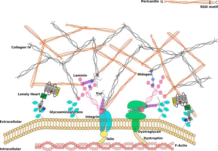
Figure 10.
Schematic representation of an extracellular matrix with incorporated Pericardin. The ECM is coupled to the actin cytoskeleton via integrins or dystroglycans. Laminins form the connection between the large structural ECM constituents, including collagen IV, Pericardin, and integrin/dystroglycan. Based on our results, we postulate that Lonely heart binds to glycosaminoglycans of membrane-associated proteins via its first speculative GAG-binding site and to Pericardin (directly or indirectly) via its second and fourth thrombospondin type 1 repeat. Loh itself is shown as a protein domain scheme with a signal peptide indicated in light blue, the ADAMTS spacer in green, the PLAC domain in yellow, and the remaining thrombospondin type 1 repeats in gray, of which two harbor a predicted GAG binding site (red bars). The form of the scheme is based on the protein model shown in Fig. 1C. The depicted interaction between Pericardin and integrin is based solely on the presence of an RGD motif at the C terminus of Pericardin and is not experimentally proven.

