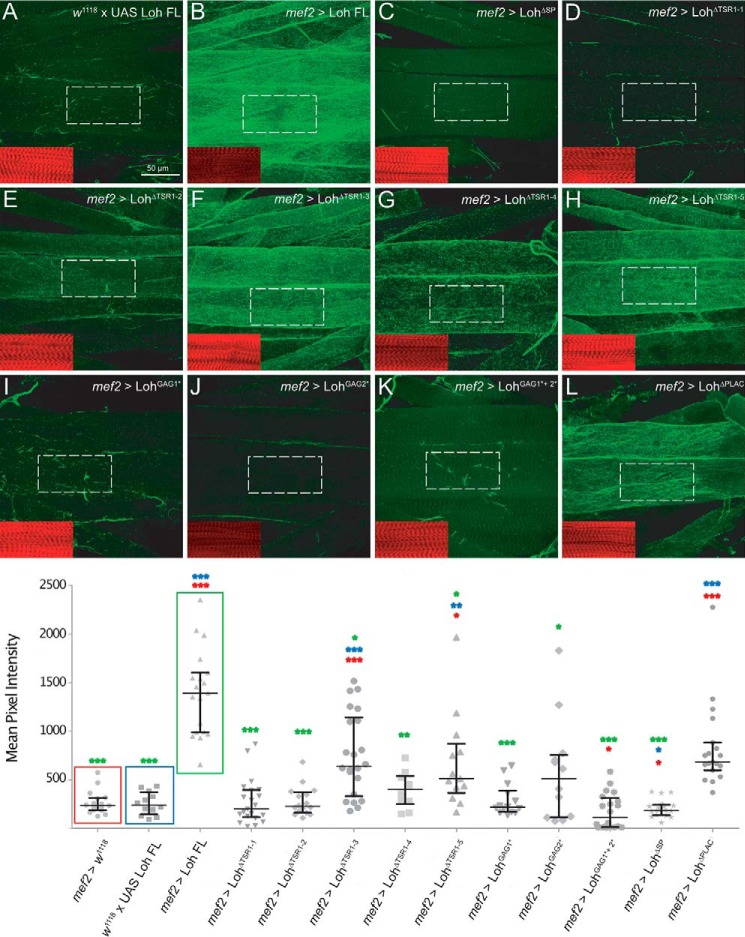
Figure 4.
Pericardin recruitment efficiency. To quantify Pericardin recruitment efficiency of individual Loh constructs, expression was driven by crossing in mef2-Gal4. Third instar larval offspring were prepared and stained for Pericardin (anti-Prc, green channel) and counterstained for F-actin (phalloidin, red channel, inset). Images were recorded with identical settings, and ROIs were measured using the “sum slices” method implemented into ImageJ. As a negative control, animals with the genotype w1118, +/UAS-Loh FL were used (A). Pixel intensity obtained for Pericardin staining (green channel) was normalized against F-actin staining (red channel). Quantification is presented as a scatter plot. Full-length Loh recruits Pericardin to the muscle matrix (B). Significant recruitment is also obvious for Loh constructs lacking the third TSR domain (UAS-LohΔTSR1–3) (F), the fifth TSR domain (UAS-LohΔTSR1–5) (H), or the PLAC domain (UAS- LohΔPLAC) (L). In Loh constructs lacking the signal peptide, no Pericardin signal above background is observed (C). Loh harboring a deleted first thrombospondin type 1 repeat (UAS-LohΔTSR1-1) (D) or a mutation of the embedded speculative GAG-binding site (UAS-LohGAG1*) (I) exhibits a considerably reduced capacity to recruit Pericardin. A strong reduction of Pericardin recruitment is also seen in LohΔTSR1-2, in LohΔTSR1-4, and in LohGAG2* mutants (E, G, and J). Loh proteins carrying mutations in both predicted GAG binding sites (LohGAG1*+2*) do also exhibit severely impaired Pericardin recruitment (K). Deleting the PLAC domain has no effect on Loh secretion on Pericardin recruitment (L). The box plot/scatter plot (bottom) depicts quantification of the construct-specific recruitment efficiencies. Colored asterisks indicate the respective significance levels (Student's t test; *, p < 0.05; **, p < 0.01; ***, p < 0.001) as calculated for the individual controls (colored boxes).

