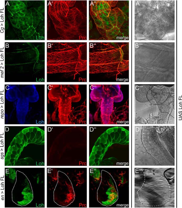
Figure 5.

Tissue-specific recruitment of Pericardin. Lonely heart was ectopically expressed in different tissues to analyze the ability of the corresponding extracellular matrices to incorporate Pericardin. Drivers used are Cg-Gal4 for adipocytes (A), mef2-Gal4 for myocytes (B), repo-Gal4 for glial cells of the central nervous system (C), sgs-Gal4 for salivary glands (D), and en-Gal4 for imaginal discs (E). Anti-Loh staining (green (A, B, D, and E) or blue (C) channel) and anti-Pericardin staining (red channel (panels labeled with a prime symbol)) are shown individually and as merged images (panels labeled with a double prime symbol). A‴, B‴, C‴, D‴, and E‴ depict bright-field images of the individual tissues. Dashed lines (E) highlight the border of the wing imaginal disc. All panels show maximum intensity projections of confocal sections. With the exception of salivary glands (D), Pericardin is recruited to all tissues tested (A, B, C, and E).
