Figure 9.
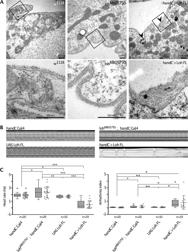
Ultrastructural and physiological effects of modulated Loh levels in cardiac tissue. A, transsections of the heart chamber of wandering third instar larvae analyzed by transmission EM. Images in the top panels represent overviews (×20,000), whereas the bottom panels depicts close-ups of the marked areas. Black arrowheads indicate ECM accumulations that mainly occur at membrane invaginations of cardiomyocytes in loh overexpression animals (handC > Loh FL) but are absent in control animals (w1118) or loh null mutants (lohMB05750). B, representative M-modes (10-s videos) of third instar larvae of the depicted genotypes indicate heart rate over time and occurrence of arrhythmia. C, quantification of B. Both heart rate and rhythmicity (arrhythmia index) are significantly affected in loh knockout animals (lohMB05750; handC-Gal4) and loh overexpression larvae (handC > Loh FL), compared with controls (handC-Gal4 and UAS-Loh FL). Depicted are mean values ± S.D. (error bars). Corresponding significance levels are indicated (asterisks, Student's t test; *, p < 0.05; **, p < 0.01; ***, p < 0.001; n.s., not significant). Data are presented as box plots and scatter plots.

