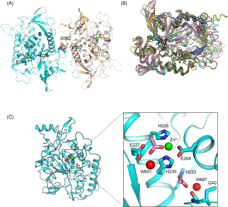
Figure 1.
Overall structure of LC/HA. (A) Cartoon representation of an LC/HA crystallographic dimer. The Zn2+- and Ca2+-coordinating residues are drawn as sticks and labeled. Ca2+ and Zn2+ are shown as silver and green spheres, respectively. (B) Structural superposition of LC/HA and the LCs of seven established BoNT serotypes and TeNT: LC/A (PDB code: 1XTF) (Breidenbach and Brunger 2004), white; LC/B (2ETF), salmon; LC/C (2QN0) (Jin et al.2007), slate; LC/D (2FPQ) (Arndt et al.2006), orange; LC/E (1T3A) (Agarwal et al.2004), yellow; LC/F1 (2A97) (Agarwal, Binz and Swaminathan 2005), magenta; LC/G (1ZB7) (Arndt et al.2005), lime; TeNT-LC (1YVG) (Rao et al.2005), forest, LC/HA, cyan. (C) The active site of LC/HA. Two acetate ions identified near the active site are drawn as pink sticks and two interacting water molecules are shown as red spheres.

