Figure 2.
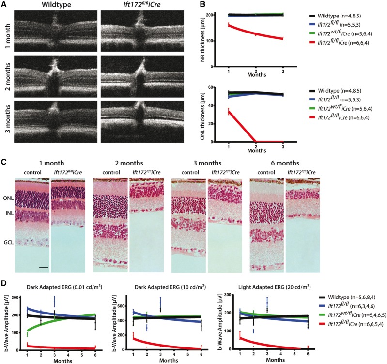
Ift172fl/fliCre mice display rapid retinal degeneration. (A) OCT data obtained at 1, 2 and 3 months of age in Ift172fl/fliCre and wild-type control mice shows diminishing thickness of ONL in the Ift172fl/fliCre mice. (B) Graphical representation of the neural retina and the ONL thickness measured from OCT images in Ift172fl/fliCre and control genotypes demonstrates decreased neural retinal thickness mainly due to thinning and eventual loss of the ONL by 2 months. (C) Representative histology sections from Ift172fl/fliCre and control mice. The scale bar represents 50 μm. (D) b-wave amplitudes from dark-adapted (0.01 and 10 cd/m2 light stimulus) and light-adapted (20 cd/m2 light stimulus) ERGs performed on Ift172fl/fliCre and control genotypes. The number of mice used in each ERG and OCT experimental condition is indicated in brackets in the order of time points taken (i.e. 1, 2, 3 and 6 months).

