Fig. 4.
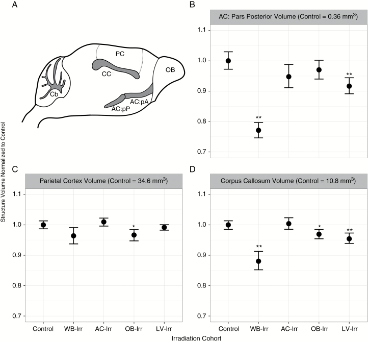
Volume differences in central and cortical brain structures after whole-brain and focal irradiations. (A) The location of the AC: pars posterior (AC:pP), parietal cortex (PC), and corpus callosum (CC) in relation to the OBs and cerebellum (Cb) are shown in a mouse brain schematic. (B–D) Brain structure volumes of the AC: pars posterior (B), parietal cortex (C), and corpus callosum (D) for all cohorts are shown. Error bars represent 95% confidence intervals; *q < 0.05 and **q < 0.01 relative to controls.

