
Fig. 3.
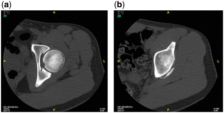
Preoperative CT scan of the left hip evaluating integrity of the posterior acetabular wall (a) and indicating mild flattening and small ossification of the posterior wall (b), which were determined to be negative for posterior wall fracture by an Orthopaedic Traumatologist.