Figure 4.
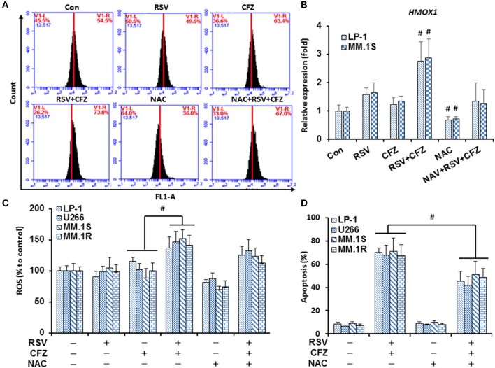
RSV and CFZ combination increased ROS production in MM cells. (A), LP-1 cells were treated with RSV (50 μM), CFZ (40 nM), or their combination treatment with or without pretreatment of NAC (3 mM, 2 h). Then, cells were harvested after 24 h for DCFH-DA staining to measure ROS production by flow cytometry. (B), LP-1 and MM.1S cells were seeded in 6-well plates and were treated the same as in (A). Cells were harvested in Trizol. Expression of HMOX1 mRNA was quantified by RT-PCR. #P < 0.01. (C), LP-1, U266, MM.1S, and MM.1R cells were treated the same as in (A). ROS production was measured by flow cytometry. P < 0.001, * compared with groups as indicated. (D), LP-1, U266, MM.1S, and MM.1R cells were treated the same as in (A). Next, cells were harvested to evaluate apoptosis through flow cytometry using FITC-Annexin V/PI staining assay. P < 0.01, * compared with groups as indicated. Data represent the mean ± SD for three separate experiments performed in triplicate.

