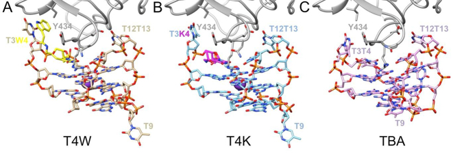
Figure 4.
Overall structures of the α-thrombin complexes with (A) T4W (PDB ID: 6EO6; this work), (B) T4K (PDB ID: 6EO7; this work) and (C) TBA (PDB ID: 1hao). Only a portion of the thrombin molecule (gray ribbon) is shown in the panels, and aptamer molecules are colored by atom, with carbon atoms of T4W, T4K and TBA colored in tan, light-blue and pink, respectively. The modified nucleotide W is highlighted in yellow, the modified nucleotide K is highlighted in magenta and selected residues are labeled.

