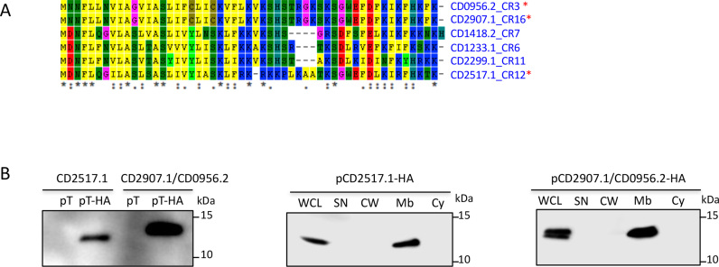
Figure 5.
Potential type I toxin proteins alignment and analysis. (A) Proteins alignment using ClustalW. ‘*’ on the right indicates toxins from three TA modules selected for detailed analysis. ‘*’ at the bottom indicates conserved residues. (B) Western-blot detection and localization of HA-tagged small proteins in the membrane fraction of C. difficile cell extracts. WCL: whole cell lysate; SN: supernatant; CW: cell wall; Mb: membrane; Cy: cytosolic fraction. Immunoblotting with anti-HA antibodies detected a major polypeptide of ∼10 kDa in whole cell lysates of the strain carrying Ptet-T(CD2517.1 or CD2907.1/CD0956.2)-HA (pT-HA) construct grown in the presence of the 250 ng/ml ATc inducer but not in extracts of strains expressing non-tagged toxins (pT) (left panel). The culture of strains carrying Ptet-T-HA plasmids induced with 250 ng/ml ATc was fractionated into cell wall (CW), membrane (Mb) and cytosolic (Cy) compartments and immunoblotted with anti-HA antibodies (middle and right panels). Proteins were separated on 12% Bis-Tris polyacrylamide gels in MES buffer.

