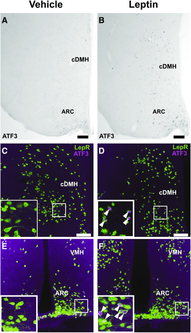
Figure 2.
ATF3 is induced specifically in LepRb neurons following acute leptin stimulation. LepRbeGFP-L10a mice were treated i.p. with vehicle or leptin (5 mg/kg i.p.) for 6 h. Brains were harvested, sectioned, and stained for ATF3 and/or GFP. Images are representative of 3–4 mice per treatment group. A and B: IHC detection of ATF3-IR (black nuclei) in the hypothalamus of control (A) and leptin-treated (B) mice. C–F: Dual IHC/immunofluorescence for ATF3 and eGFP in the DMH (C and D) and ARC (E and F) of control (C and E) and leptin-treated (D and F) LepRbeGFP-L10a mice. White arrowheads indicate colocalized neurons. Scale bars = 100 μmol/L. cDMH, caudal DMH; VMH, ventromedial hypothalamus.

